Neurological problemes

MUSCULOSKELETAL AND NEUROLOGICAL DISEASES
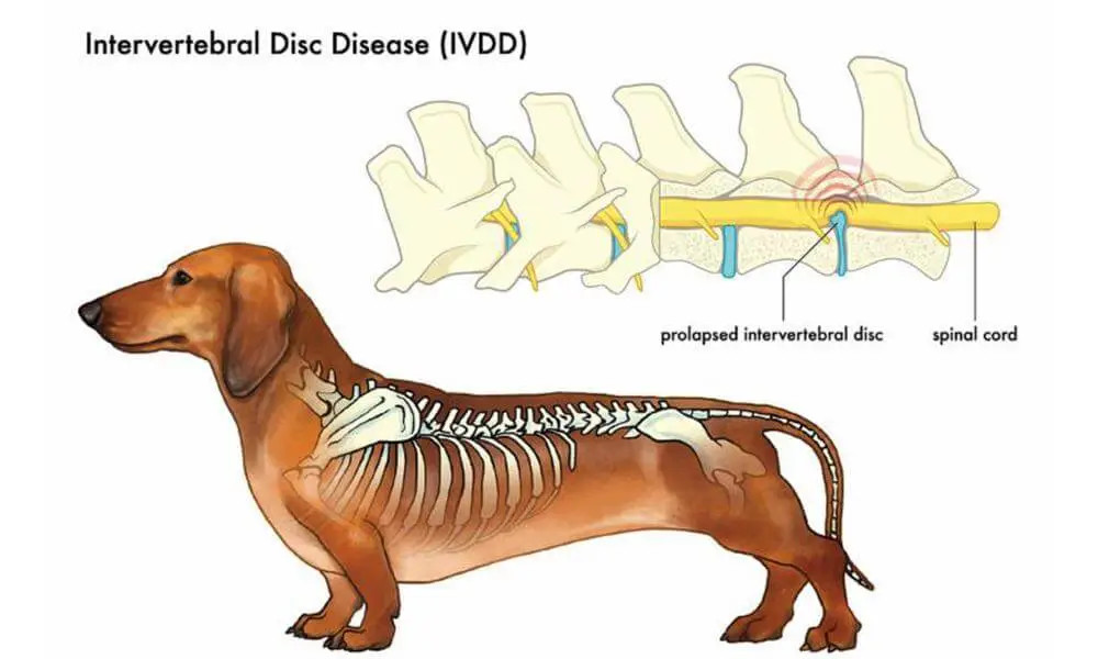
Intravertebral Disc Disease (Herniated Disc)

Introduction:
Even our four-legged friends can develop intervertebral disc disease, which is known in two main forms. In one case, spinal injuries caused by a trauma (e.g. car accident) can trigger sudden palsy symptoms in an otherwise healthy pet. The second (and more common) form occurs when the elasticity and water content are slowly lost from the disc and it rotates toward the spinal canal, eventually causing herniation and sudden paralytic symptoms. Most disc ruptures occur in the mid to low back, but can also occur in the neck region.
Symptoms:
Dorsal herniation can cause a humpbacked appearance, and possible limping or clumsiness of the hind limbs. It will also cause severe pain, hind limb paralysis, or bladder bloating and consequent urination issues. Contracted back and abdominal muscles as well as skin hypersensitivity may also be seen. In the case of cervical herniation, neck and forelimb issues will arise.
Diagnosis:
To diagnose a herniated disc, thorough physical and neurological examination, radiographs with spinal cord contrast (myelograms), and MRIs may be used.
EPILEPSY

Introduction:
Epileptic symptoms are caused by abnormal functioning of the cortical brain. The cortex can cause a disease called primary, or true, epilepsy. However, other organ systems (e.g. kidney, liver) can also be affected which is referred to as secondary epilepsy.
Symptoms:
Epileptic symptoms are very similar no matter the cause, usually seem to occur without any reason, and they typically last for the same amount of time. Symptoms are largely dependent on cortical localization. In milder cases there may be behavioral disorders, motor dysfunction, or seizing muscle groups (e.g. involuntary lip movements, chewing). In severe cases, confusion, loss of consciousness or even muscle spasms all over the body can be seen.
Diagnosis:
A video diary of the epileptic symptoms greatly help with diagnosis and development of an effective treatment by identifying the cortical area causing epilepsy. In the case of primary epileptic animals, after the seizure occurs, only an EEG-test can be used to determine any abnormality. Secondary epilepsy diagnosis may include blood sampling or spinal fluid testing. Brain morphological changes can be detected by CT and MRI.
Treatment:
It is important to distinguish true epilepsy as is often a ghostly similar to the seizures sometimes caused by panic attacks. Antiepileptics should be given only if the frequency and severity of the attacks requires.
